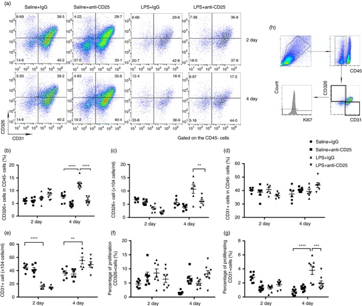
Figure 3.

Pulmonary epithelium is impaired and endothelial proliferation is abrogated in regulatory T‐cell (Treg)‐depleted mice. (a) Cytofluorometric dot plots of epithelial and endothelial cells within days after injury. Numbers depict the fraction of CD45‐ cells within the designated gate. (b) Summary data for the percentage and (c) number of CD326 + epithelial cells in the lungs depicted in (a). (d) Summary data for the percentage and (e) number of CD31 + endothelial cells in the lungs depicted in (a). (f) Percentage of proliferating CD326 + cells and (g) percentage of proliferating CD31 + cells in the lungs. (h) Schematic gating of ki67 + epithelium or endothelium. n = 6, Mean ± SEM. *P ≤ 0·05; **P ≤ 0·01; ***P ≤ 0·001; and ****P ≤ 0·0001.
