Fig. 1.
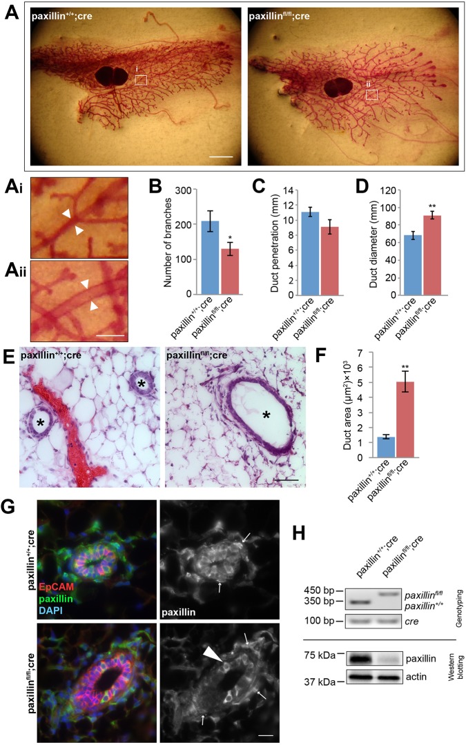
Paxillinfl/flcre mammary glands have fewer branches and dilated ducts. (A) Whole-mount staining of 6-week-old paxillin+/+;cre and paxillinfl/fl;cre mammary glands. Scale bar: 2 mm. (Ai,Aii) Higher-magnification images showing that paxillinfl/fl;cre have larger duct diameters (arrowheads). Scale bar: 250 μm. (B) Quantification of the total number of branches in the mammary gland, n=10 animals (paxillin+/+;cre), n=13 (paxillinfl/fl;cre). (C) Quantification of duct penetration. n=6 (paxillin+/+;cre), n=8 (paxillinfl/fl;cre). (D) Quantification of duct diameter, n=6 (paxillin+/+;cre), n=7 (paxillinfl/fl;cre). (E) Hematoxylin and Eosin staining of 6-week-old mammary gland sections. Asterisks indicate individual ducts. Scale bar: 50 μm. (F) Quantification of duct cross-section area, n=3 (paxillin+/+;cre), n=5 (paxillinfl/fl;cre). (G) Paxillin expression pattern in 6-week-old mammary glands. Occasional mosaic expression of paxillin was observed in paxillinfl/fl;cre glands (arrowhead). Arrows indicate myoepithelial cells. Scale bar: 20 μm. (H) Genotyping from mice tail snips and western blotting of paxillin using lysates obtained from mammary gland extract. Student's t-test was performed. Data are mean±s.e.m. **P<0.01.

