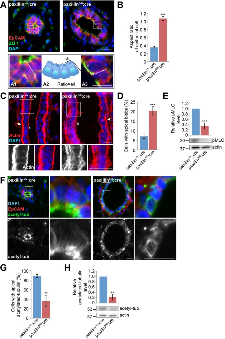
Fig. 2.
Paxillinfl/flcre mammary glands have disrupted epithelial cell shape and disorganized cytoskeletal components. (A) Six-week-old mammary gland sections labeled for the tight junction marker ZO1 and the baso-lateral membrane marker EpCAM. Higher magnification of the boxed areas are shown in (A1) paxillin+/+;cre and (A3) paxillinfl/fl;cre. (A2) Schematic of the apical and lateral cell membrane measurements used for calculating ratios. Scale bars: 20 μm. (B) Quantification of apical-to-lateral membrane length ratio, n=3 (at least 20 cells per animal). (C) Actin staining of longitudinal sections of mammary gland ducts. Asterisks indicate the apical surface; arrows indicate the myoepithelial layer. Scale bars: 20 μm. (D) Quantification of the number of cells with apical F-actin blebs, n=6 (paxillin+/+;cre), n=7 (paxillinfl/fl;cre) (at least three ducts per animal). (E) Mammary epithelial cell lysates blotted for pMLC and quantification of the relative level, normalized to actin. (F) Six-week-old mammary gland stained for acetylated tubulin. Scale bars: 20 μm. (G) Quantification of cells with apical acetylated tubulin, n=5 (paxillin+/+;cre), n=4 (paxillinfl/fl;cre) (at least 28 cells per animal). (H) Mammary epithelial cell lysates blotted for acetylated tubulin and quantification of the relative expression level. Student's t-test was performed. Data are mean±s.e.m. **P<0.01, ***P<0.001.

