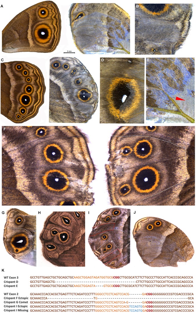
Fig. 2.
Crispants generated by targeting exon 2 and exon 3 of Dll. (A) Wild-type forewing of B. anynana (left) and exon 3 phenotype with eyespots missing in areas of lighter pigmentation and disrupted venation (right). (B) Exon 3 phenotype with missing eyespot in a patch with mutant clones. (C) Wild-type hindwing of B. anynana (left) and exon 3 phenotype with split eyespots and bisected eyespot centers. (D) Exon 3 phenotype showing light colored scales in mutant clones across an eyespot. (E) Exon 3 phenotype showing close-up of crispant in A with a region of missing scales as indicated by the red arrowhead. (F-J) Exon 2 mutations. (F) Wild-type (left) and crispant wing (right) of the same individual in which ectopic eyespots appeared on the distal hindwing margin after exon 2 was targeted. (G) Comet-shaped Cu1 eyespot center. (H) Example of a spontaneous comet mutant. (I) Wing with ectopic eyespots as well as missing eyespots. (J) Missing eyespots on hindwing in mosaic areas also showing lighter pigmentation. (K) Next-generation sequencing of selected crispants (exon 3 top panel and exon 2 bottom panel) identifying the most frequent indels around the target site. Orange, guide region; red, PAM sequence; blue, insertions; dashed lines, deletions. Dotted lines on exon 2 crispants in F, G and I represent wing regions carefully dissected for DNA isolation. Wing sectors for the crispant shown in I, outlined in red (missing eyespots), were pooled for DNA isolation as were wing sectors outlined in white (ectopic eyespots). For the exon 3 crispant in C the entire distal wing margin was dissected and for the crispant in D the area around the eyespot was dissected.

