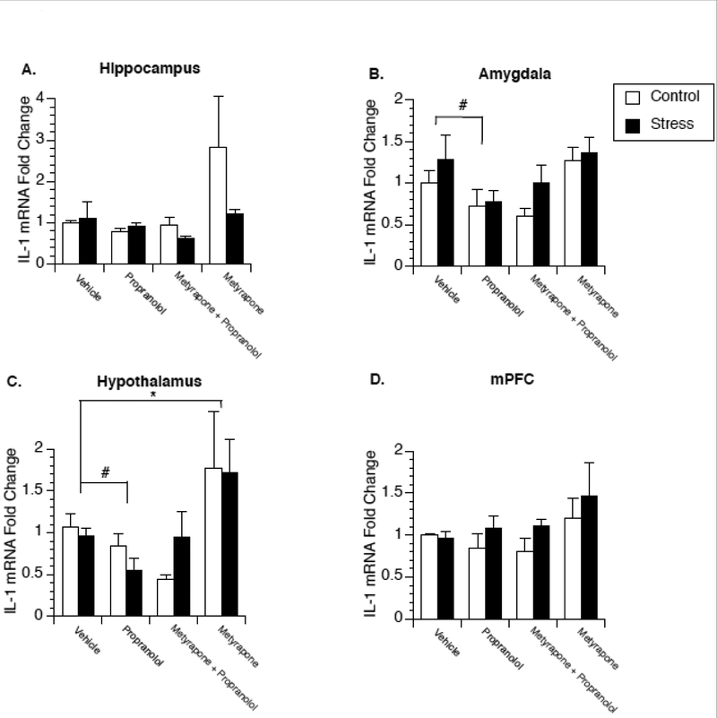
Figure 3.
Effect of stress on glucocorticoid and β-adrenergic receptor regulation of brain IL-1β expression in female rats. Rats were exposed to 4 days of stress or remained in their home cage to serve as controls. Twenty-four hours after the last stressor animals were administered either saline, propranolol, metyrapone, or a combination of metyrapone and propranolol and two hours later euthanized for measurement of IL-1β mRNA in the hippocampus (A), amygdala (B), hypothalamus (C), and medial prefrontal cortex (D). A significant main effect of propranolol was observed in the amygdala and hypothalamus. A significant main effect of metyrapone was observed in the hypothalamus. * represent significant (p < 0.05) main effect of metyrapone treatment compared to saline-injected animals. # represent significant (p < 0.05) main effect of propranolol treatment compared to saline-injected animals. Values represent mean IL-1β mRNA fold change compared to non-stressed, vehicle treated animals. Symbols and bars represent group mean ± SEM.

