
Figure 6.
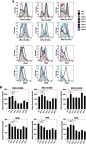
Flow cytometry of activation markers on BMDCs. BMDCs were incubated with each type of the liposomes (fc: 50 μM) for 2 hrs and then washed off. After further incubation overnight in the fresh culture medium, the cells were labeled with fluorescent antibodies against MHC-I (H-2Db), MHC-I (H-2Kb), MHC-II (I-A/I-E), CD80, CD86, and CD40 followed by flow cytometry (A, see gating strategies in Figure S6). (B) The mean fluorescent intensities were measured and data show the mean + SD of two independent experiments with two replicates, respectively. *P<0.05, **P<0.01. Note that the liposomes were not associated with OVA. LPS was used as a positive stimulus. Abbreviations: BMDCs, bone marrow-derived dendritic cells; LPS, lipopolysaccharide; OVA, ovalbumin.