
Figure 7.
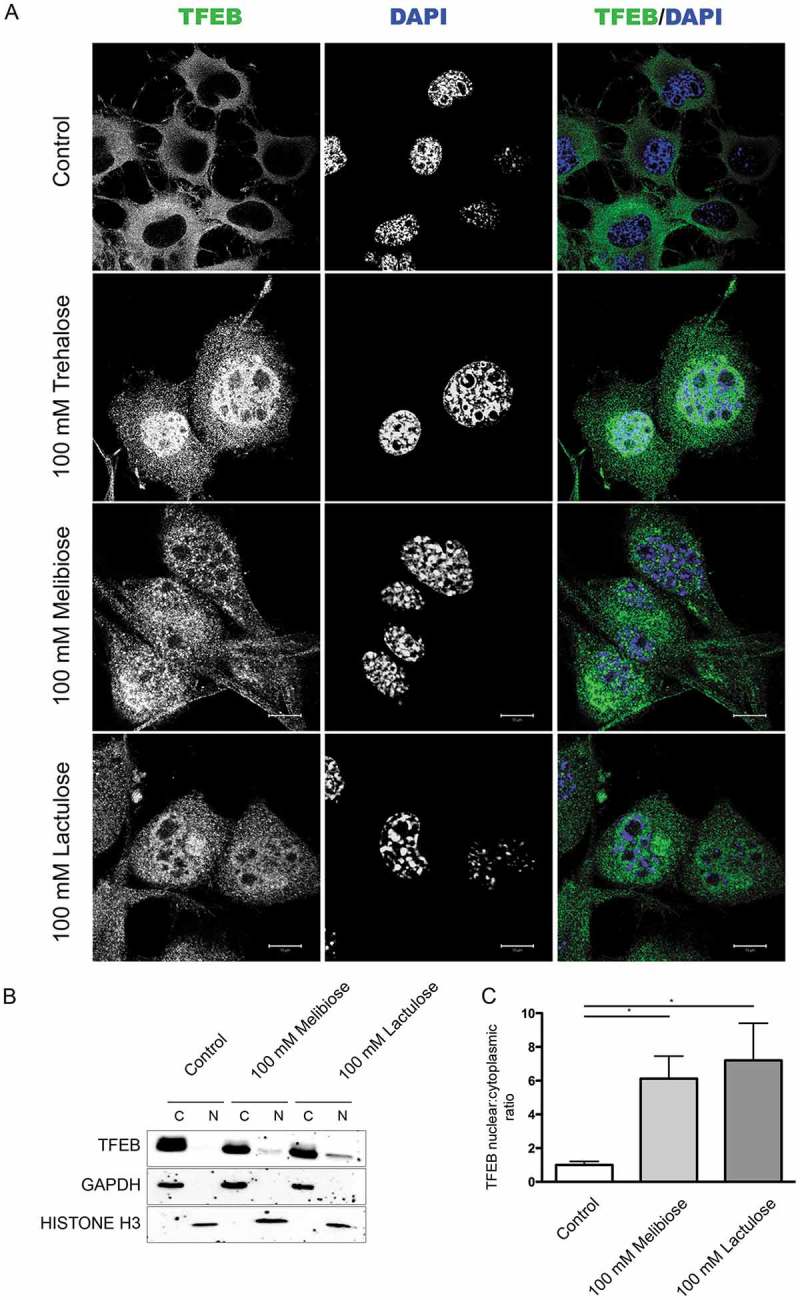
Melibiose and lactulose induce TFEB nuclear translocation. (a) IF analysis of TFEB localization performed on NSC34 cells treated with 100 mM trehalose, 100 mM melibiose or 100 mM lactulose for 48 h. Nuclei were stained with DAPI (blue) (63X magnification). Scale bar: 10 μm. (b) WB analysis of cytoplasmic (C) and nuclear extracts (N) on NSC34 cells untreated (control) or treated with 100 mM melibiose or 100 mM lactulose for 48 h. GAPDH and histone H3 were used as internal loading control for cytoplasmic and nuclear fractions, respectively. (c) The bar graph represents the mean ± SD for n = 4 independent samples of nuclear:cytoplasmic TFEB ratio compared to untreated cells (*p < 0.05 one-way ANOVA with Tukey’s test).