
Figure 5.
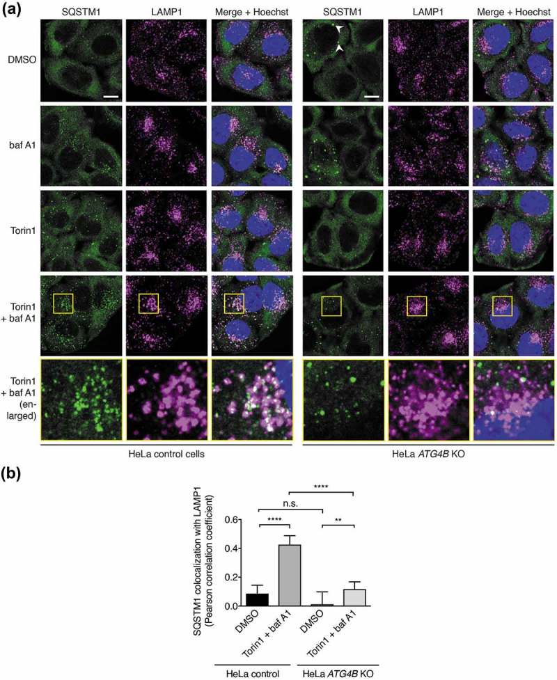
Impaired autophagic delivery of SQSTM1 to lysosomes in cells lacking ATG4B. (a) Immunocytochemistry of endogenous SQSTM1 and LAMP1 in control and ATG4B KO HeLa cells. Cells were treated for 3 h with DMSO or 250 nM Torin1 + 10 nM baf A1 prior to fixation and staining. White arrowheads indicate examples of large SQSTM1 bodies in ATG4B KO cells that likely correspond to protein aggregates. Yellow boxes show region-of-interest in the Torin1 + baf A1 treated condition that is enlarged in the panels below. Scale bar: 10 µm. (b) Assessment of colocalization between endogenous SQSTM1 and LAMP1 in HeLa control and ATG4B KO cells using single-plane confocal images as represented in Figure 5(a). Bars indicate mean and error bars show standard deviation (n = 10 randomly-selected cells per condition). **** P ≤ 0.0001, ** P ≤ 0.01, n.s. (not significant) P > 0.05 (Sidak’s multiple comparison test).