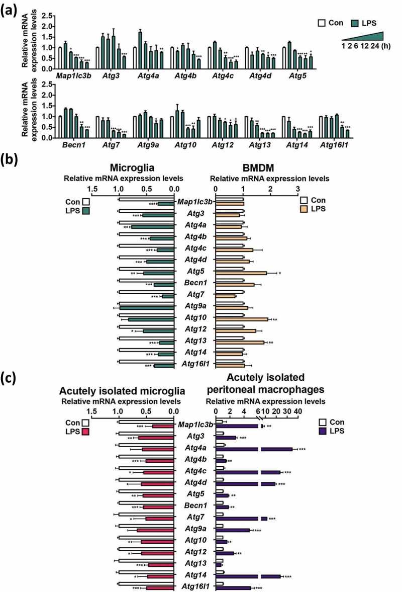
Figure 2.

LPS decreases the expression of Atg genes in primary microglia. (a) Time course analysis of the mRNA levels of Atg genes by qRT-PCR following LPS (1 μg/mL) treatment (n = 3). (b) Comparison of relative mRNA levels of Atg genes between primary microglia (n = 5) and BMDMs (n = 4) 24 h after LPS (1 μg/mL) treatment. (c) Comparison of relative mRNA levels of Atg genes, between microglia and peritoneal macrophages acutely isolated from the same LPS-injected mice (5 mg/kg, intraperitoneally), 24 h post-injection (n = 3). In all experiments, mRNA levels were normalized to Actb. All data are mean ± SEM. *P < 0.05, **P < 0.01, and ***P < 0.001 compared to the control (Con).
