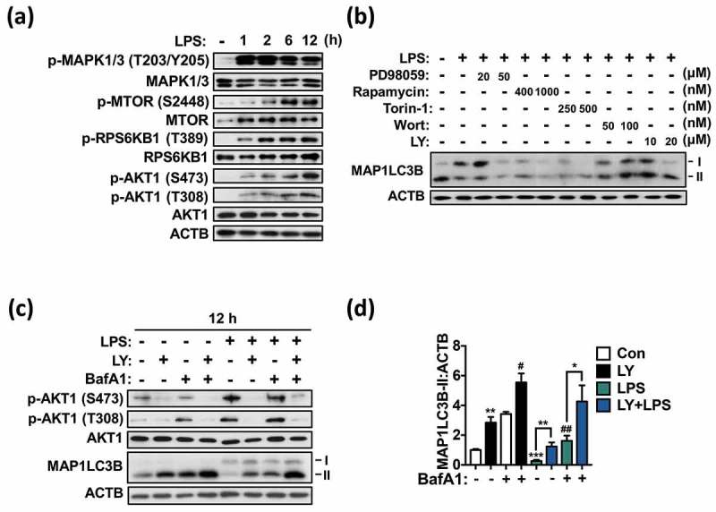
Figure 6.

LPS suppresses autophagy via the PI3K-AKT1 signaling pathway in primary microglia. (a) Western blotting analyses of phosphorylation of AKT1 (T308, S473), MTOR (S2448), MAPK1/3 (T203/Y205), and RPS6KB1 (T389) following LPS treatment (1 μg/mL) in primary microglia. (b) Western blotting analysis of MAP1LC3B-II level after inhibition of MAPK1/3 (with PD98059), MTOR (with rapamycin or Torin-1) or PI3K (with LY294002; LY or wortmannin; Wort) in primary microglia treated with LPS (1 μg/mL) for 12 h. (c) Analysis of autophagic flux after inhibition of PI3K with LY (20 μM) in primary microglia treated with LPS (1 μg/mL) for 12 h. (d) Quantitative analysis of MAP1LC3B-II levels in primary microglia. In all experiments, the blots shown are representative of at least 3 experiments with similar results. All inhibitors were added 1 h prior to LPS treatment. All data are mean ± SEM. *P < 0.05, **P < 0.01, and ***P < 0.001 compared to the control (Con) unless indicated otherwise. #P < 0.05 and ##P < 0.01 compared to the control treated with BafA1 only.
