
Figure 1.
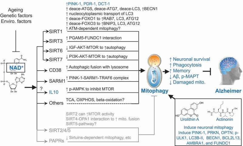
Schematic representation of how NAD+, urolithin A, and actinonin induce mitophagy, and inhibit Alzheimer disease (AD). Cellular NAD+ levels are reduced with ageing as well as affected by genetic and environmental (enviro.) factors. NAD+ is a cofactor of sirtuins (SIRT1 to SIRT7), CD38, SARM1, and PARPs. The nuclear SIRT1, SIRT6, SIRT7, mitochondrial SIRT3, CD38, and SARM1 induce mitophagy/autophagy. NAD+ may induce mitophagy through other pathways such as through the induction of the mitophagy inducer IL10 and fundamental metabolic pathways. Increased NAD+ may also inhibit autophagy/mitophagy through cytoplasmic SIRT2, mitochondrial SIRT4, mitochondrial SIRT5, and the DNA damage sensor PARPs. One reasonable explanation is that a robust NAD+-dependent mitophagy induction and a mild NAD+-dependent mitophagy inhibition give an outcome of a remaining robust induced mitophagy. UA and AC are robust mitophagy inducers, both inducing expression of mitophagy/autophagy proteins such as PINK1, PRKN, OPTN, p-ULK1, LC3B-II, BECN1, BCL2L13, AMBRA1, and FUNDC1. Results marked in blue are from the current study. Results marked in dark (induction of mitophagy) and gray (inhibition of mitophagy) are from previous publications. See manuscript for details and references. deace, deacetylated.