
Figure 4.
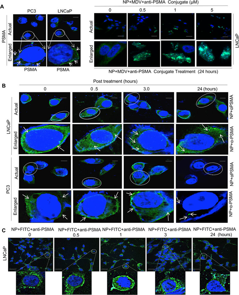
PSMA specific internalization of hybrid nanoparticle in prostate cancer cells. (A) Immunolocalization of prostate specific membrane antigen (PSMA) in PSMA proficient and PSMA deficient prostate cancer isogenic LNCaP and PC3 models (left panel) and expression of PSMA in LNCaP cells in response to varying doses of hybrid nanoparticle-loaded enzalutamide–anti-PSMSA conjugates with indicated concentrations (right panel). (B) Confocal microscopic images of anti-PSMA internalization via PSMA in PSMA proficient and deficient LNCaP and PC3 isogenic PCa lines with indicated time points. (C) Confocal microscopic images of FITC in FITC-loaded (FITC load mimics MDV3100 payload) hybrid nanoparticle–anti-PSMA conjugates in LNCaP cells with indicated time points. Each data point is a mean ± SD from three or more independent experiments. ★★p < 0.05 was considered as statistically significant. Scale bar “–” = 20 μm.