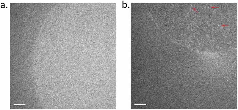
Figure 3.

Fluorescence of hPiezo1-GFP in DHBs. (a) Autofluorescence from a DPhPC bilayer with interior consisting of empty azolectin liposomes. (b) Fluorescence of hPiezo1-GFP inserted in bilayer diffusing throughout the bilayer. Example puncta indicated with red arrows. Diffusion can be seen more clearly in Supplementary Movies 1 & 2. Exposure time: 100 ms; Laser power: 0.2 µW/um2; Image brightness scaled to range from 300–1600 a.u, on Andor EMCCD Camera (see Methods). Scale bar indicates 10 um.
