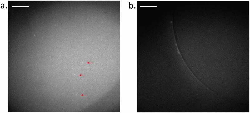
Figure 4.

Fluorescence of hP1-GFP in DHBs with 8% cholesterol. (a) hP1-GFP diffusing in a bilayer composed of 8:92 cholesterol to DPhPC (see Methods). Example puncta are indicated by red arrows. (b) Positive control for fluorescence, but not membrane protein insertion, observed using a DPhPC bilayer with an interior of GFP-filled azolectin liposomes (fluorophore present, but no membrane protein). Background fluorescence is observable but not individual puncta diffusing inside the bilayer. Diffusion can be seen more clearly in Supplementary Movies 3 & 4. Exposure time: 100 ms; Laser power: 0.3 µW/um2; Image brightness scaled to range from 150–300 a.u on Hamamatsu 95B camera. Scale bar indicates 20 µm.
