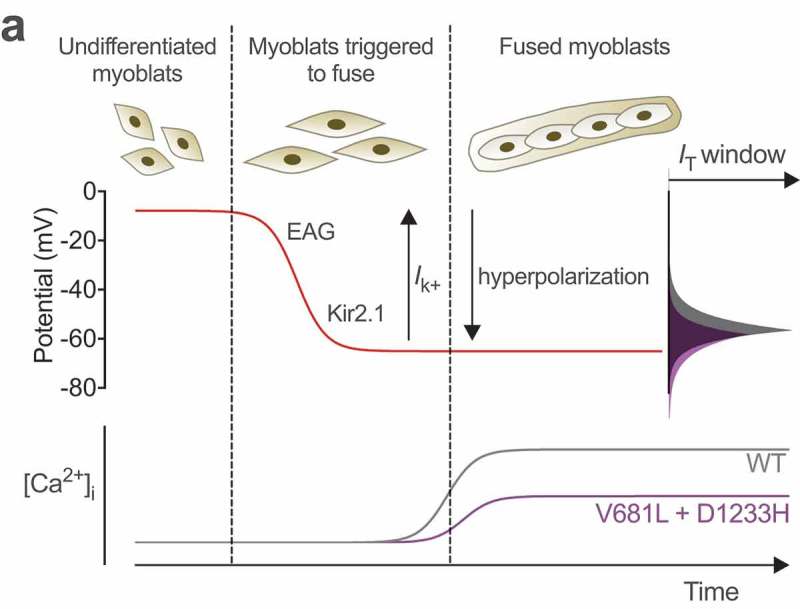
Figure 5.

Hypothetical model for altered myogenesis by Cav3.2 variants. While the membrane potential of undifferentiated myoblast is about −8 mV, sequential expression of the slow-inactivating voltage-gated ether-a-go-go (EAG) and inward-rectifying Kir2.1 potassium channels brings the membrane potential to approximately −65 mV (red line), allowing window calcium influx through Cav3.2 channels (grey line) required for the fusion of myoblasts. Reduced window current caused the by V681L and D1233H mutations results in a decreased window calcium influx (purple line) which potentially compromises early stage myogenesis. Adapted from [30].
