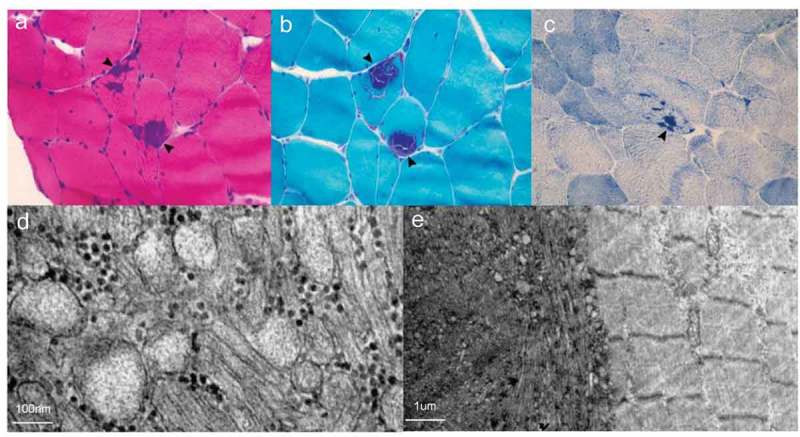
Figure 6.

Muscle biopsy of Case 2.
HE (A), MGT (B) and NADH-TR (C) demonstrates tubular aggregates (arrowhead) and vacuoles (arrow) under the light microscope. D. Tubular aggregates under electron microscopy. E. The right area shows relatively normal skeletal muscle structure and the left area shows tubular aggregates under electron microscopy.
