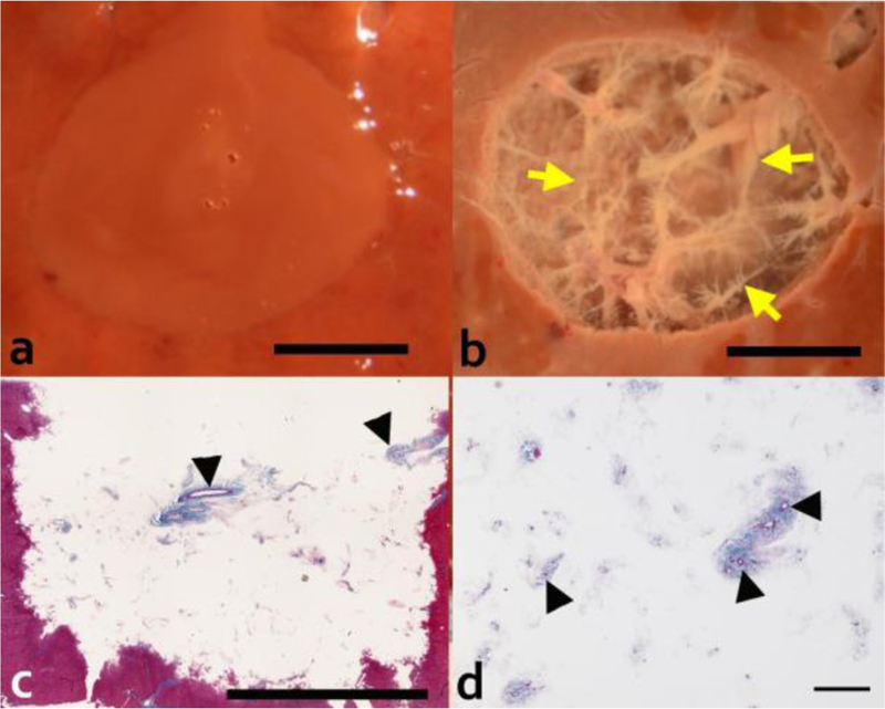
Figure 5.

Gross and histological evaluation of the 2 cm diameter volumetric lesion generated with 1 ms pulses at 600 W peak acoustic power and 0.4% dc. Upper row show photos of lesions in cross-section (a) before and (b) after rinsing the contents out. Rinsed lesions reveal connective tissue (yellow arrow) that was not liquefied by the treatment. Masson’s trichrome stained histological sections of the rinsed lesion (longitudinal orientation) (c) confirm the presence of connective tissue appearing in the native state throughout the depth of the lesion. Scale bar represents 1 cm. (d) Higher magnification of the lesion with the homogenized contents washed out reveals intact fibrillar collagen with patent small caliber vessels and ducts (black arrow head). Scale bar represents 250 μm.
