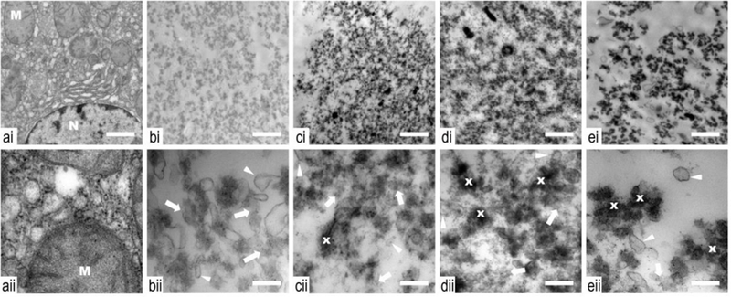
Figure 7.

Transmission electron microscopy images of samples taken from control (a) and histotripsy lesions produced with a range of duty cycles: 1 (b), 3 (c), 5 (d) and 10% (e), obtained at low (i) and high (ii) magnifications. Untreated tissue (a) shows a generally normal appearance with an intact nucleus (N) with disperse chromatin. A mild general edema is present as evident by slightly swollen mitochondria (M) with some loss of membrane cristae, which can be expected in ex vivo tissue. With treatment, and at all duty cycles, all cellular structure is lost. The cytoplasmic cell membranes are absent and cellular components are not distinguishable. Ghosts of organelles can be observed in the form of membranous structures (white arrow head) of variable size and electron density amongst a sea of cellular debris. With increasing duty cycle, there is evidence of a gradient increase in thermal application. The cellular debris become more condensed (x) and electron dense with increasing duty factor, consistent with heat coagulation of proteins. Non coagulated proteins are observed in a higher percent at the lower duty factors (white arrow). Low (i) and high (ii) magnification scale bar represents 1 μm and 250 nm, respectively.
