Figure 1.
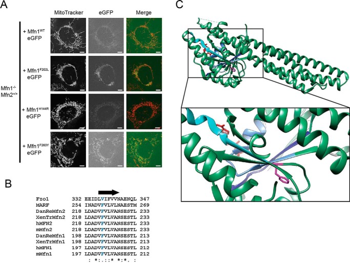
Mfn1 Phe-202 is a highly conserved residue in a central β strand. A, representative images of Mfn1−/− Mfn2+/+ cells stably expressing Mfn1-eGFP or Mfn1F202L-eGFP following retroviral transduction. Mitochondria were labeled with MitoTracker Red CMXRos and visualized by fluorescence microscopy. Images represent maximum intensity projections. Scale bars are 5 μm. B, sequence alignment of Mfn1 F202 region generated using Clustal Omega. The arrow indicates the position of β strand 4G, and the conserved phenylalanine (Mfn1 Phe-202 and Mfn2 Phe-223) is in blue text. C, Mfn1IM crystal structure with Phe-202 highlighted in fuchsia. Critical residues in conserved catalytic domains are highlighted: the P-loop in cornflower blue, G2/switch I in light blue, G3/switch II in dark blue, and G4 in turquoise (Protein Data Bank (PDB) code 5GOE).

