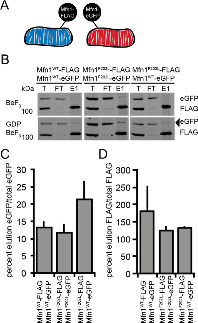
Figure 6.

Tethering assay to assess the physical interaction of Mitofusin proteins in trans. A, schematic of the differential epitope labeling utilized in the tethering assay. B, mitochondria were isolated from a clonal population of Mfn1WT-FLAG-expressing Mfn1-null cells (Mfn1WT Mfn2+/+), a clonal population of Mfn1WTeGFP-expressing Mfn1-null cells (Mfn1WT Mfn2+/+), a clonal population of Mfn1F202LFLAG-expressing Mfn1-null cells (Mfn1F202L Mfn2+/+), and a clonal population of Mfn1F202L-eGFP-expressing Mfn1-null cells (Mfn1F202L Mfn2+/+) and incubated with BeF3 in the absence or presence of GDP. Following lysis, immunoprecipitation was performed with α-FLAG magnetic beads. Proteins eluted from the beads were subjected to SDS-PAGE and immunoblotted with α-Mfn1. The arrowhead indicates the eGFP protein eluted from FLAG beads. Total (T) represents 3% of the input, flow-through (FT) represents 3% of the unbound protein, and elution (E) represents 40% of the immunoprecipitated protein. C, the percentage of the indicated Mitofusin-eGFP in the elution compared with the total is shown as the mean +S.D. of three independent experiments. D, the percentage of the indicated Mitofusin-FLAG in the elution compared with the total is shown as the mean + S.D. of three independent experiments.
