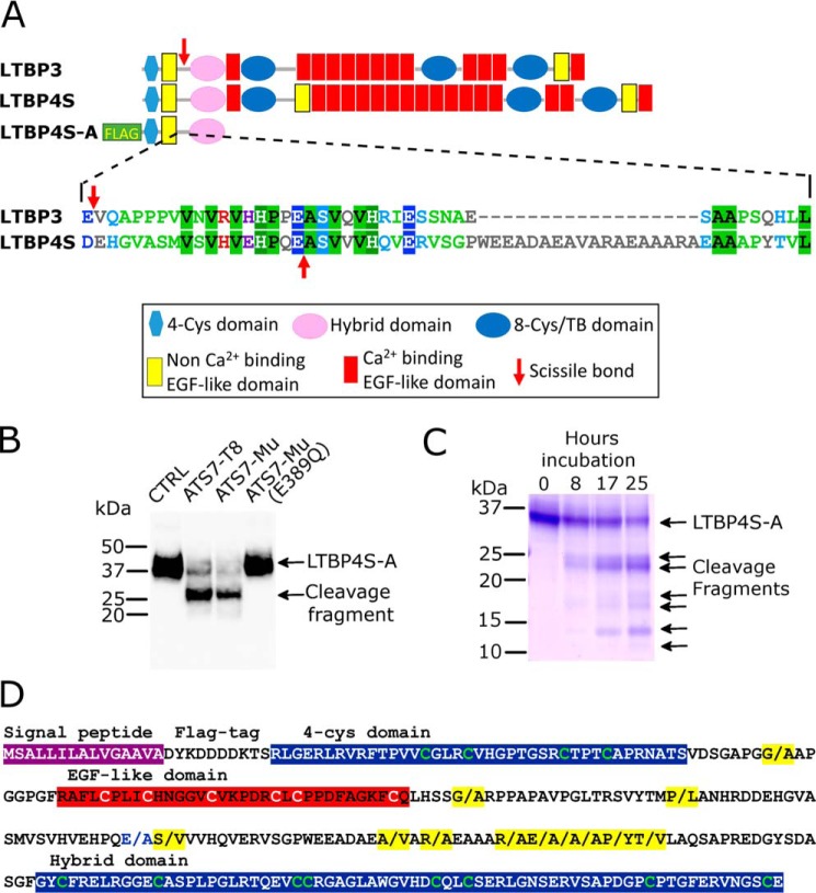
Figure 4.
Identification of LTBP4 as a substrate of ADAMTS7. A, domain organization of LTBP3 and LTBP4S, which is the shorter isoform of LTBP4, indicating the location of the scissile bonds (red arrow) that were identified by TAILS. LTBP4S-A is a truncated recombinant variant that contains an N-terminal FLAG-tag. For the linker region that contains the identified scissile bonds, an alignment of the amino acid sequence of LTBP3 and LTBP4 is shown, indicating the scissile bonds (red arrows). B, Western blotting of LTBP4S-A conditioned media (anti-FLAG Ab) incubated for 17 h with buffer (CTRL), purified ADAMTS7-T8 (40 nm), ADAMTS7-Mu conditioned medium, or ADAMTS7-Mu(E389Q) conditioned medium. C, Coomassie Blue–stained SDS-PAGE gel of purified LTBP4S-A (10 μm) incubated with 10 nm purified ADAMTS7-T8 for 0, 8, 17, and 25 h. D, amino acid sequence of LTBP4S-A highlighting scissile bonds cleaved by ADAMTS7 (indicated by/and highlighted in yellow) as identified by LC-MS/MS analysis following TMT labeling of cleaved and uncleaved LTBP4S-A.

