Figure 8.
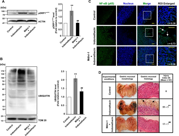
Mdivi-1 prevents indomethacin-induced mitochondrial damage, inflammation, and gastric injury. Immunoblot analyses for DRPSer-616 phosphorylation (A) and mitochondrial proteome ubiquitination (B) in control, indomethacin-treated and Mdivi-1 + indomethacin-treated rat gastric mucosa. Actin and TOM20 were used as the loading control for total cell lysates and mitochondrial fractions respectively. Numerical values at the side of the blots indicate the positions of corresponding molecular mass markers. Bar graphs adjacent to the blots represent the densitometric analyses of the immunoblot data after normalization with the respective loading controls. C, immunohistochemical analysis of nuclear translocation of NF-κB upon indomethacin treatment or Mdvi-1 + indomethacin treatment; white arrows indicate mucosal epithelial cells with higher degree of nuclear NF-κB. NF-κB (green) was immunostained by anti-NF-κB (p65) primary and Alexa Fluor 488-tagged anti-rabbit secondary antibodies. Nucleus (blue) was stained by DAPI. Scale bars correspond to 50 μm. Corresponding values in the inset represent Pearson's correlation coefficient (r) of the blue and green signals corresponding to nucleus and NF-κB, respectively. Random tissue sections were screened, and a representative image of one of the several experiments has been presented. D, gastric mucosal morphology and histology by hematoxylin–eosin staining. Black arrows indicate injured mucosal surface. For all animal experiments n = 6–8, and experiments were performed three times. **, p < 0.01 versus control, and ##, p < 0.01 versus indomethacin as calculated by ANOVA followed by Bonferroni's post hoc test. A detail of each method is described under “Experimental procedures.”

