
Figure 3.
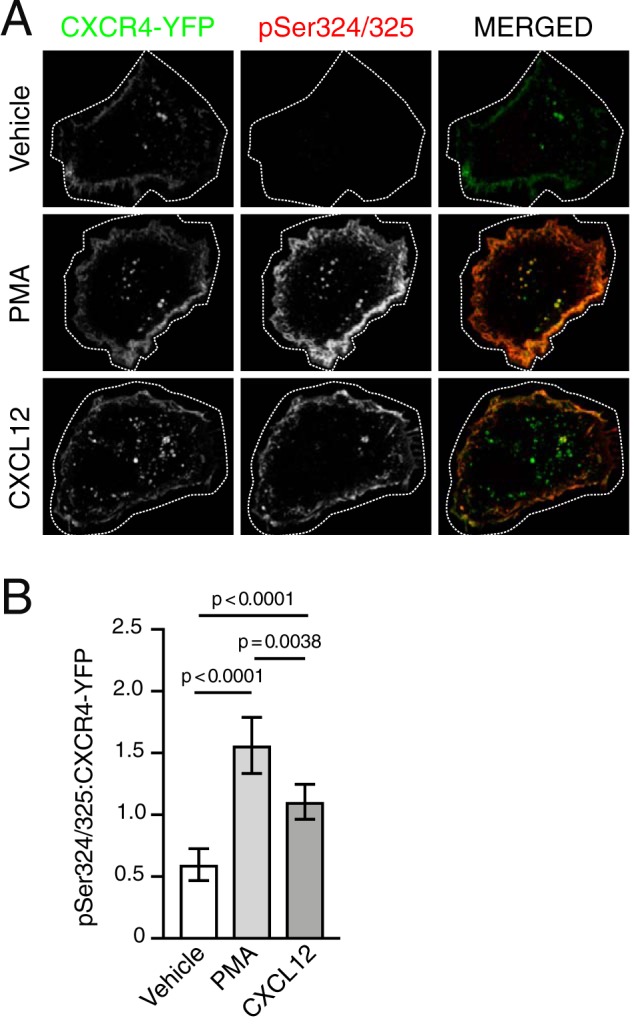
PKC is sufficient to mediate site-specific C-tail phosphorylation of CXCR4. A, representative micrographs of HEK293 cells transiently expressing HA-CXCR4-YFP and immunostained to detect simultaneous phosphorylation of serine residues 324 and 325 (pSer324/325). Cells were serum-starved for 3.5 h then stimulated with either vehicle, 10 nm PMA, or 10 nm CXCL12 for 30 min. Panels show HA-CXCR4-YFP (left) and pSer324/325 (center) as single-channel grayscale and pseudocolored, merged images (right) for the indicated treatment conditions. Images were processed in ImageJ, as described under “Experimental procedures.” For optimal viewing, HA-CXCR4-YFP and pSer324/325 signal intensities were adjusted with opacity screens in Photoshop. Screens for HA-CXCR-YFP were adjusted to offset between group variation in expression; screens for pSer324/325 were adjusted within each group to match adjustments in HA-CXCR4-YFP. Scale bar, 10 μm. B, quantification of pSer324/325:HA-CXCR4-YFP fluorescence intensities. Bars represent the average from three independent experiments. Error bars, 95.00% CI. For vehicle, n = 13 cells; for PMA, n = 15 cells; for CXCL12, n = 17 cells. Data are presented as mean pSer324/325:HA-CXCR4-YFP fluorescence intensity ratios. Data were analyzed using Welch's ANOVA followed by Tamhane's T2 multiple-comparison test. Adjusted p values are indicated.