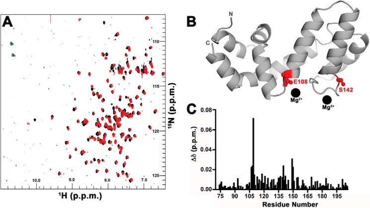
Figure 5.
RGS17 binds Mg2+ in solution. A, 1H-15N 2D HSQC spectra of RGS17 alone (black) or upon addition of 15 mm MgCl2 (red). B, structure of RGS17 where residues with CSPs greater than 0.15 ppm are shown in ball-and-stick in red. In contrast to spectra obtained in the presence of CaCl2, MgCl2 induces CSPs in two regions of RGS17. The locations of the Mg2+ ions (black spheres) are modeled based on the location of Ca2+ atoms observed in the RGS17–Ca2+ crystal structure. C, graph of CSPs for all the residues that could be assigned in the 1H-15N 2D HSQC spectra for RGS17.

