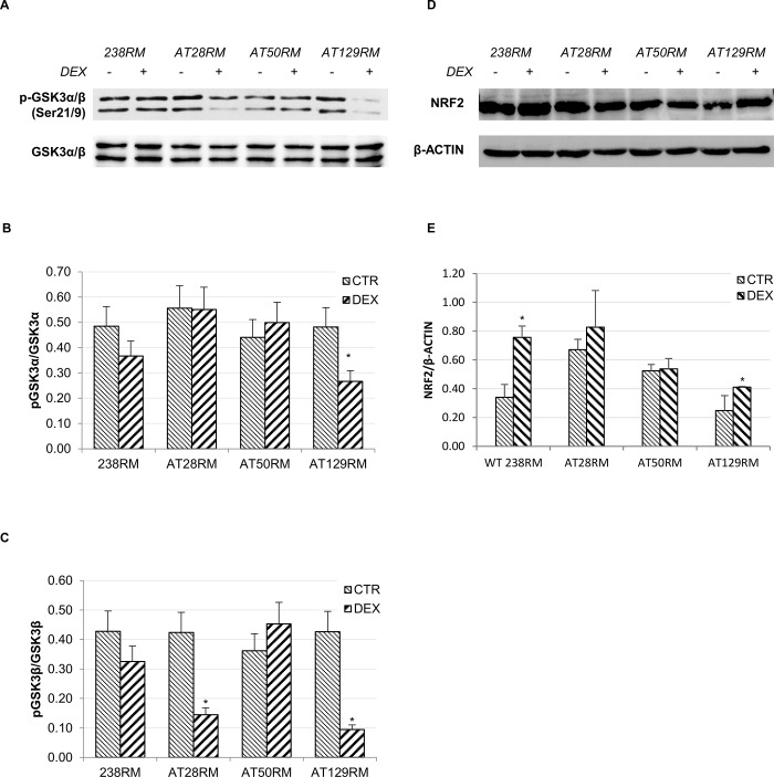
Fig 3. GSK3 is not inhibited by DEX in AT cells.
The activation status of the inhibitory kinase GSK3 was investigated in WT and AT cells. A) Western blot analysis for phosphorylated GSK3-α/β and total GSK3-α/β, in WT (238RM) and AT (AT28RM, AT50RM, AT129RM) cells treated as reported in Fig 2. B),C) Quantification of p-GSK3-α/total GSK3-α and of p-GSK3-β/total GSK3-β ratio, respectively. D) Western blot analysis for total NRF2 protein levels in the four cell lines used in this study, treated as in Fig 2. β-ACTIN served as a loading control. E) Quantification of the relative amounts of NRF2 in the total cell extracts of WT and AT cells shown in D. Blots shown are representative and the histograms are the means and SEM of four independent experiments. (Wilcoxon signed rand test; *two-tailed p-values<0.05).

