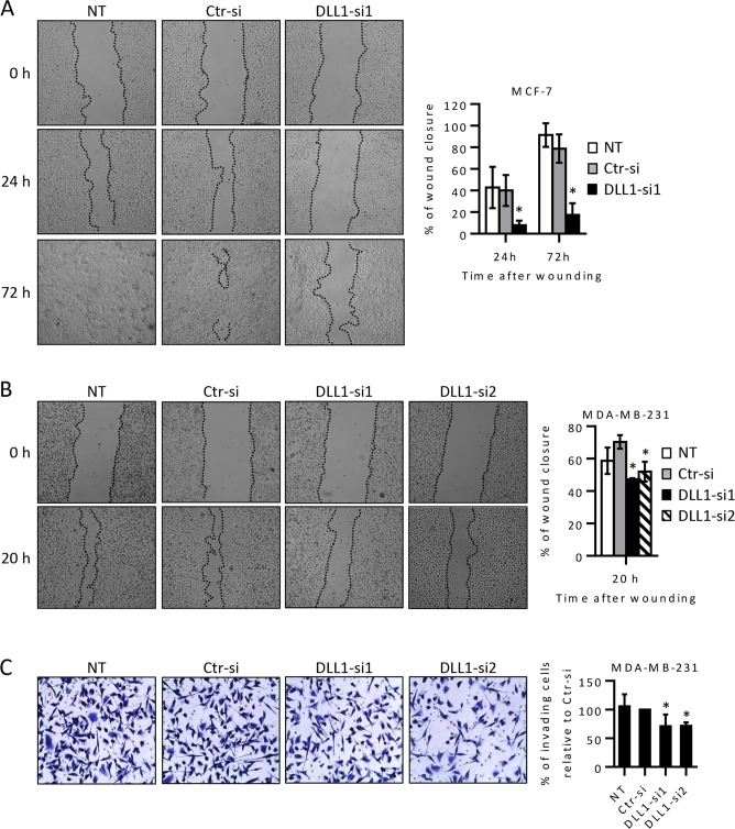
Fig 2. DLL1 downregulation decreases migration of MCF-7 and MDA-MB-231 cells and the invasive potential of MDA-MB-231 cells.
MCF-7 and MDA-MB-231 cells were transiently transfected with DLL1 siRNAs (DLL1-si1/2), negative control siRNA (Ctr-si) or not-transfected (NT) as indicated. (A-B) At 55–70 hours after transfection, cells at 80–90% confluency were scratched, and wound closure was evaluated by microscopy at various time points. Representative images taken at the indicated times post-wounding from three independent experiments are shown. The graph represents mean percentage values (+ SD) of wound closure at each analyzed time point from scratches of these assays. (C) MDA-MB-231 cells were collected 72 hours after transfection and equal cell numbers were added to the upper chamber of 8-μm-pore membranes coated with matrigel and their invasion was measured. Representative fields of crystal-violet-stained cells that invaded to the lower surface of the membranes are shown in each condition. The graphs show mean percentage values (± SD) of invading cells of three independent experiments. *, P < 0.05, compared with Ctr-siRNA transfected cells.

