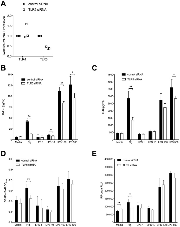
Fig 5. TNF-α and IL-8 production but not NF-κB or IRF3 activity induced by LPS in THP1 cells is impaired upon silencing of TLR5.
THP1-Dual Cells were differentiated with Vitamin D3 and plated at 50,000 cells per well were transfected for 48 hours with a negative control siRNA or with siRNA against TLR5 at 5 nM. Cells were unstimulated or stimulated overnight with either E. coli O111:B4 LPS at varying doses in ng/ml or recombinant B. pseudomallei flagellin (Flg) at 100 ng/ml as a positive control. A: TLR4 and TLR5 mRNA expression were measured by quantitative RT-PCR in cell lysates after 48 hrs of transfection and are expressed relative to ubiquitin C (UBC). Data from three independent experiments are shown, normalized for each experiment. TNF-α (B) and IL-8 (C) concentrations were determined in supernatants the day following stimulation. Transcription factor activation was determined using (D) SEAP-NF-κB and (E) IRF-Lucia luciferase reporter assays, expressed as OD630nm and relative light units, respectively. B-E: Data are displayed as mean ± standard deviations of quadruplicate conditions; one representative example of two or three independent experiments is shown. *, P<0.05; **, P≤0.01.

