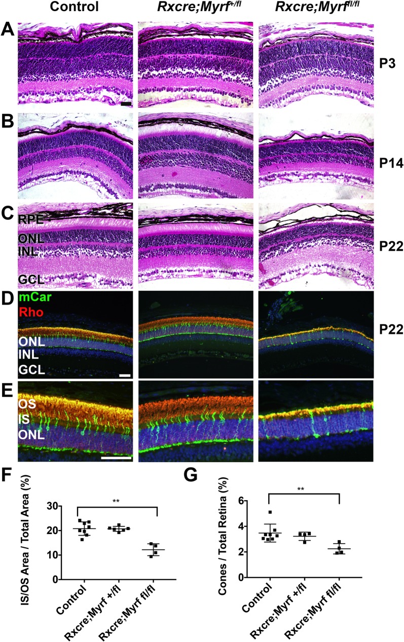
Fig 6. Loss of MYRF leads to early retinal degeneration in mice.
(A-C) H&E histology of P3 (A), P14 (B), and P22 (C) control (Myrf+/fl or Myrffl/fl) or Myrf heterozygous and homozygous conditional knockout mice (RxCre;Myrf+/fl and RxCre;Myrffl/fl). RxCre;Myrffl/fl retinas have shortened inner and outer segments, but retinal structure during development is preserved. RxCre;Myrf+/fl are structurally indistinguishable from control. (D-E) Low magnification (D) and high magnification (E) images of photoreceptor immunolabeling in P22 animals. Mouse cone arrestin (mCar, green) and rhodopsin (Rho, red) and DAPI (blue) were used to mark cones, rods, and nuclei, respectively. (F) Quantitative analysis of inner/outer segment area compared to total retinal area. (G) Quantitative analysis of cone fraction compared to fraction of total retinal cells. There is a significant decrease in IS/OS area and cone fraction in RxCre;Myrffl/fl retinas. n = 8 eyes, 6 animals (control); n = 6 eyes, 3 animals (RxCre;Myrf+/fl), n = 4 eyes, 4 animals (RxCre;Myrffl/fl). Mean±standard deviation are plotted along with each individual eye data point. **, p<0.01. Scale bar, 50μm.

