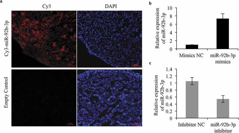
Figure 2.

Validation of efficiency of ovarian transfection of microRNA in vitro. (a) Ovaries were collected after incubation with miR-92b-3p labeled with Cy3 and evaluated by confocal microscopy. Strong red fluorescence was observed after 96 h of transfection. DAPI was used for staining nuclei. Bar = 50 μm. (b, c) Real-time PCR analysis of miR-92b-3p expression in ovaries transfected with miR-92b-3p inhibitor, miR-92b-3p mimics, or their respective controls. *p < 0.05.
