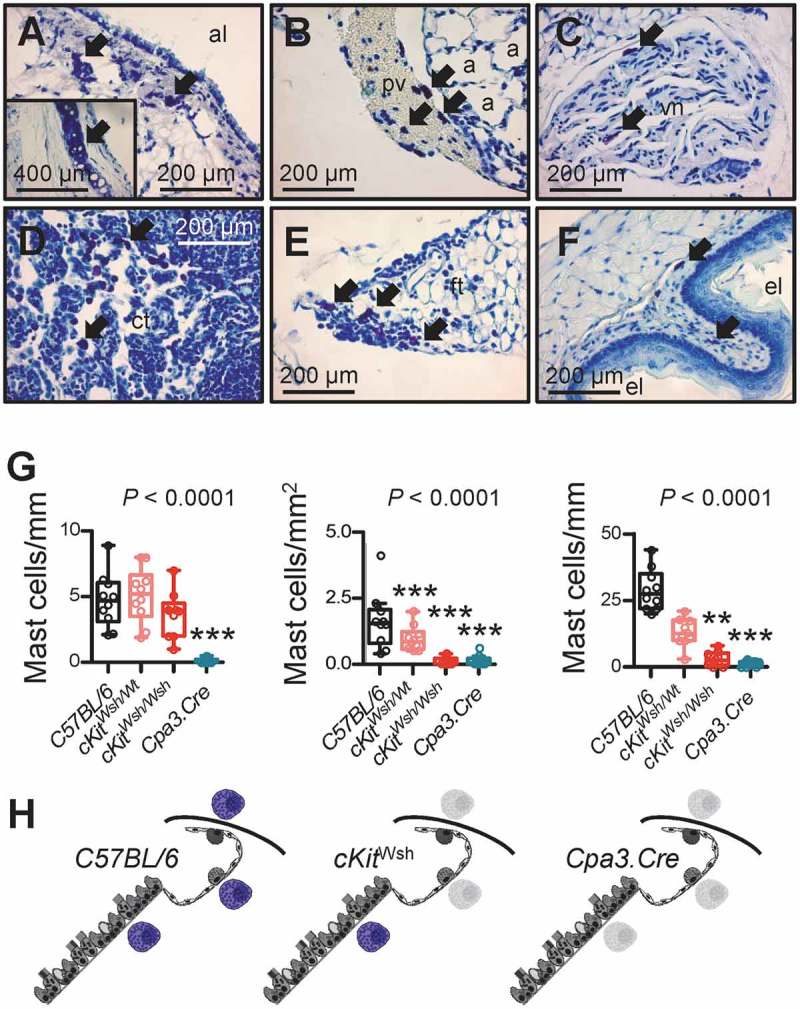
Figure 2.

Thoracic and skin mast cells in two different mouse models of mast cell deficiency.
The airways, alveoli, and skin of mice carrying one or two cKitWsh alleles (designated cKitWsh/Wt and cKitWsh/Wsh, respectively) or one Cpa3.Cre allele on a pure C57BL/6 background, and C57BL/6 littermate or Lyz2.Cre heterozygous control mice (n = 10/group) were sectioned and stained with toluidine blue. Representative microscopic images of toluidine blue-stained tissue sections (A-F), summary of data from n = 10 mice/group (G), and schematics of mast cell competence (colored mast cells) and deficiency (grey mast cell shadows) (H). A-F Arrows indicate mast cells in the submucosa of a large airway (A; inlay shows tracheal cartilage as positive control of metachromatic purple staining), in a large pulmonary vein (B), in the vagus nerve (C), in the thymus of a 6-week-old (D) and a 20-week-old (E) mouse, and in the esophageal submucosa (F) of C57BL/6 controls. a, alveoli; pv, pulmonary vein; al, airway lumen; vn, vagus nerve; ct, cellular thymus; ft, fatty thymus; el, esophagus lumen. G Airway, alveolar, and skin mast cell density of C57BL/6 control, cKitWsh/Wt, cKitWsh/Wsh, and Cpa3.Cre mice (summary of data from n = 10 mice/group). Shown are median with Tukey’s whiskers (boxes: interquartile range; bars: 50% extreme quartiles), raw data points (dots), and Kruskal–Wallis analysis of variance (ANOVA) probability (P) value. **, and ***: P< 0.01 and P< 0.001, respectively, for comparisons with C57BL/6 controls by Dunn’s post-tests. Only statistically significant differences are indicated. Note the airway mast cell competence of cKitWsh/Wt and cKitWsh/Wsh mice, and the complete mast cell deficiency of Cpa3.Cre mice. H Schematics depicting mast cells (purple) of the airways, alveoli, and skin of C57BL/6, cKitWsh/Wt, cKitWsh/Wsh, and Cpa3.Cre mice. Grey mast cell fadeouts indicate mast cell deficiency of the given anatomic compartment.
