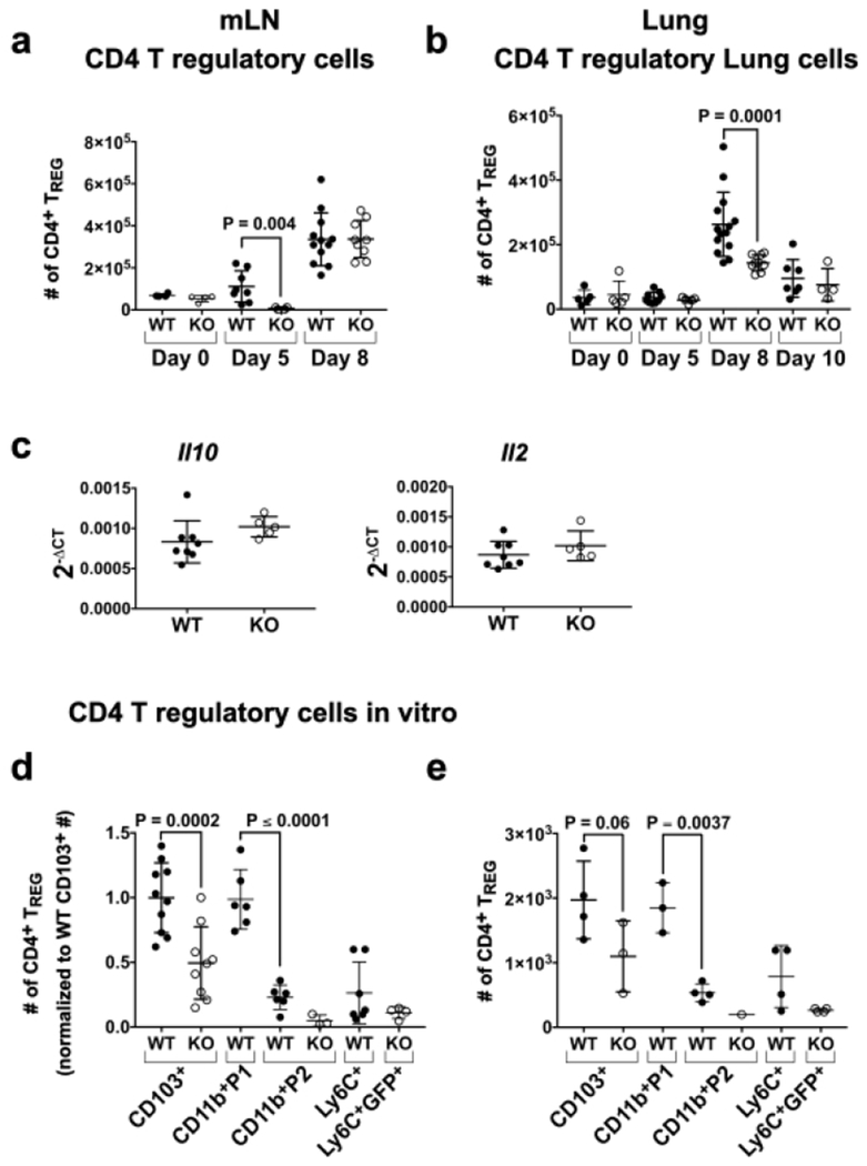
Fig. 5. CD11c-cre-Irf4f/f mice show reduced numbers of CD4+ FOXP3+ TREGs in the mLN and lung after IAV infection.
(a-b) Lung and mLN cells were harvested from infected mice on days 0, 5, 8 and 10 p.i. Surface and intracellular staining identified TREGs (Fig. S4a). Shown are numbers of CD4+CD25+FOXP3+ TREGs in the (a) mLN and (b) lung, (c) Expression of Il10 and Il2 RNA in total mLN cells on day 8 p.i. determined by qPCR. (d) Numbers of FOXP3+ TREGs in cultures normalized to the average of WT CD103+mice (gated as in Fig. S4b) after in vitro incubation of naïve CD4+ OT-II T cells with OVA323-339 peptide and isolated lung DC subsets (1000 per well) sorted on day 3 p.i. (e) Numbers of FOXP3+ TREGs in cultures from one representative experiment. Symbols represent individual mice, with the mean and SD indicated. The data are compiled from 1-3 independent experiments each with 3-5 animals per WT or KO group. Significance was evaluated using a Mann-Whitney test (panels a-c) or one-way ANOVA with Tukey’s multiple comparison test (panel d-e), with p values indicated.

