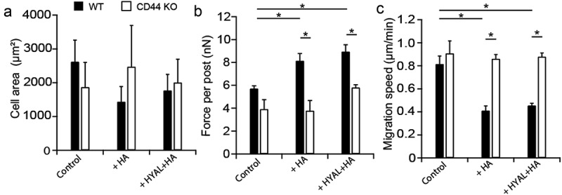
Figure 4.

CD44 KO cells migrated faster despite reduced contractility. (a) Cell spread area remained unaffected as a result of exogenous HA or a sequential exposure to HYAL+HA in both serum-starved WT (black bars) and CD44 KO (white bars) fibroblast areas. (b) For PDL WT cells, traction forces significantly increased in the presence of both exogenous HA (P = 0.026) or the combination of HYAL+HA (P = 0.026) compared to the untreated WT. However, CD44 KO cells were unaffected by the presence of exogenous HA in either conditions. Finally, no statistical difference was observed in traction force between CD44 KO and WT cells. However, both CD44 KO cells treated with exogenous HA (P = 0.025) and the sequential combination of HYAL+HA (P = 8.03e-3) appeared significantly less contractile than their WT counterparts. (c) For migration, PDL WT cells in the presence of exogenous HA (P = 0.026) or the sequential combination of HYAL+HA (P = 0.029) appeared less migratory than the WT control cells. However, CD44 KO cells appeared significantly more migratory regardless of exogenous stimulation with HA or HYAL+HA. Furthermore, when comparing CD44 KO to WT cells, we found no statistical difference in migration speed between CD44 KO cells compared to WT. However, CD44 KO cells treated with exogenous HA (P = 0.022) or the sequential combination of HYAL+HA (P = 0.010) appeared more migratory than their WT counterparts. Data shown as average ± SEM from three experimental replicates (Table S1). Asterisk indicates P < 0.05 (one-way ANOVA with a Bonferroni’s and Fischer post-hoc adjustment).
