Fig. 5.
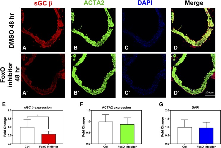
sGC expression in ex vivo murine aortas treated with FoxO inhibitor was decreased. Representative staining for ex vivo murine aortas treated with 10 μM FoxO inhibitor for 48 hours showing sGC β protein (A and A′), ACTA2 (B and B′), DAPI (C and C′), and merged channels (D and D′). Quantification of immunostaining for sGC β protein (E), ACTA2 protein (F), or DAPI staining (G). n = 3 animals. Student’s unpaired t test was used for determination of significance. *P < 0.05. Error bars represent S.D.

