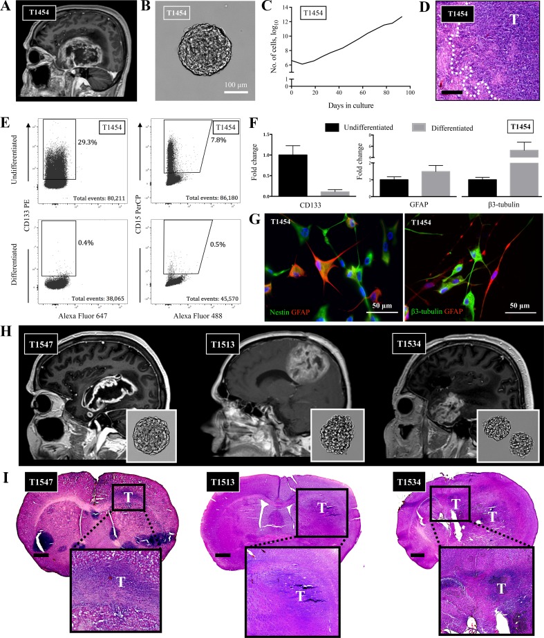
Fig. 1.
Patient-derived GSCs harbor stem cell properties. a T1 contrast-enhanced MRI of T1454 displaying the GBM located in the right temporal region. b Upon cultivation, the tumor formed free-floating spheres, c which could be exponentially propagated in serial passages. d Upon xenografting, the tumor formed an invasive tumor. Invasive rim of the tumor delineated. Staining with hematoxylin and eosin. Scale bar = 200 μm. e The GSCs in T1454 expressed the stem cell markers CD133 and CD15. The expression was reduced upon differentiation. f qRT-PCR confirmed the reduction of stem cell-related expression of CD133, along with the increased expression of the more lineage-specific GFAP and β3-tubulin upon differentiation. The results are presented as mean and standard error to the mean of three independent experiments. g Upon differentiation in serum-containing media, the tumor cells formed arborizations and twisting processes associated with a more mature morphology, and stained positive for GFAP and β3-tubulin. Nuclei stained with DAPI. h, i T1 contrast-enhanced MRI of the primary GBM T1547 and the pretreated and recurrent GBMs T1513 and T1534 with the corresponding in vitro spheroid morphology upon cultivation and the corresponding xenograft. Brain sections are stained with hematoxylin & eosin. Scale bar 1 mm

