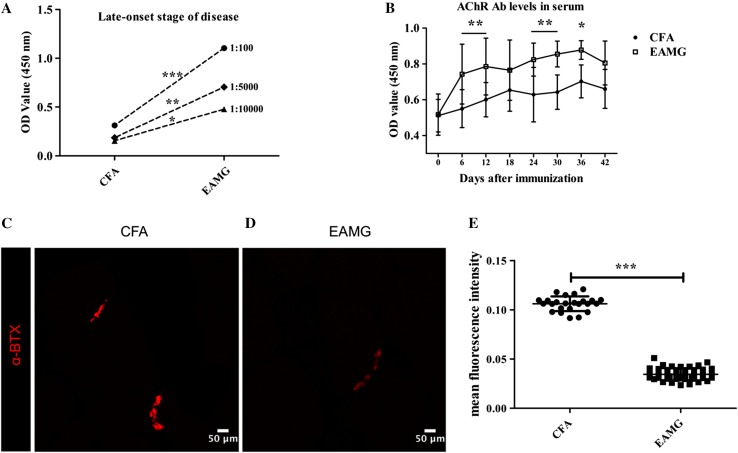
Fig. 2.
Elevated levels of auto-AChR antibodies in serum and a reduction in AChRs in muscles. A ELISAs were used to detect anti-AChR IgG titers on day 45 in samples diluted 1:100, 1:5000, and 1:10000 from the EAMG and CFA groups (n = 3 rats/group). B Anti-AChR antibody levels in serum samples collected from the two groups every 6 days after the first immunization (n = 6 rats/group) assessed by ELISA. C, D Forelimb muscle from EAMG (D) and CFA (C) rats stained with α-BTX as a marker of AChRs. Scale bars, 50 μm; n = 4 rats/group. E Fluorescence intensity values (mean ± SD; *P < 0.05, **P < 0.01, ***P < 0.001).

