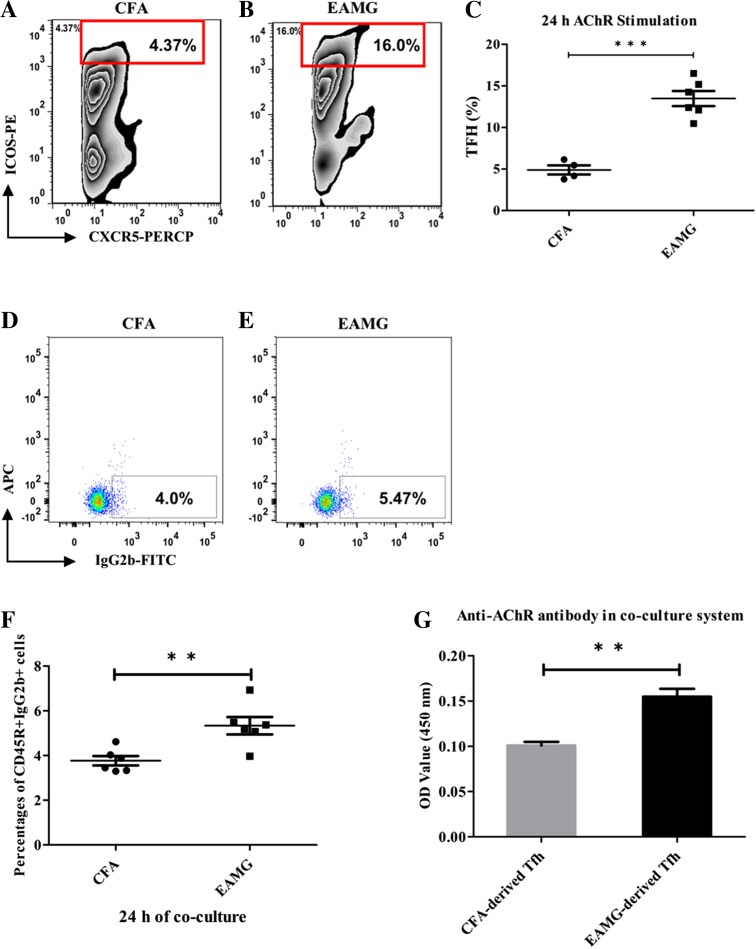
Fig. 5.
Elevated anti-AChR IgG levels in B cells co-cultured with Tfh cells. A, B The ratio of AChR-specific CD4+CXCR5+ICOS++ Tfh cells in the EAMG samples was 3–4-fold that in the CFA samples as detected by flow cytometry (C; ***P < 0.001). More IgG2b-secreting B cells were found in EAMG rats (E) than in CFA rats (D, F; ***P < 0.001) after 24 h of co-incubation with AChR-stimulated Tfh cells and the level of anti-AChR antibodies in the supernatant (G; **P < 0.01) was also elevated in the EAMG group. Data are from two independent experiments with 4–6 rats per condition per experiment and are expressed as the mean ± SD (**P < 0.01).

