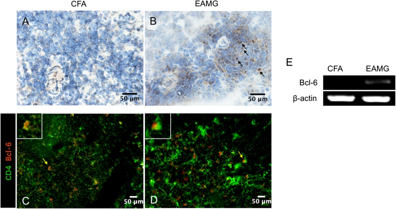
Fig. 6.
Bcl-6 expression in lymph nodes. A, B Immunohistochemical staining was used to detect Bcl-6-positive cells in lymphoid tissues from CFA (A) and EAMG (B) rats (scale bars, 50 μm; arrows indicate positive cells). C, D Immunofluorescence assays were used to detect cells positively stained for CD4 (green) and Bcl-6 (red) (Tfh cells) in lymphoid tissue (scale bars, 50 μm; arrows indicate positive cells which are further magnified in the white boxes). E Bcl-6 and β-actin mRNA levels were assessed by semi-quantitative RT-PCR in CD4+ T cells that were isolated and purified from lymph nodes from both groups. Data are from two independent experiments with 3–4 rats per condition per experiment.

