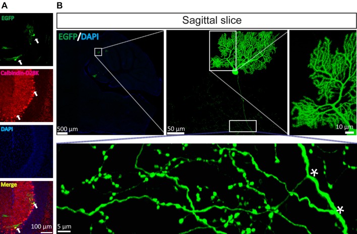
Fig. 4.
Mutant SFV VLP rapidly and sparsely labels Purkinje neurons in vivo. To sparsely label Purkinje cells, the mutant virus was injected into the third cerebellar lobule (3Cb) region. The mice were sacrificed at 24 hpi and sagittal and coronal sections were prepared. A The EGFP signals were co-localized with the Purkinje cell marker calbindin-D28k (arrows). B The fine structure of the Purkinje cells was apparent, including the dendritic tree, long axon, and axonal branches (asterisks). These images are representative of 15 sections (n = 3 mice).

