Figure 1.
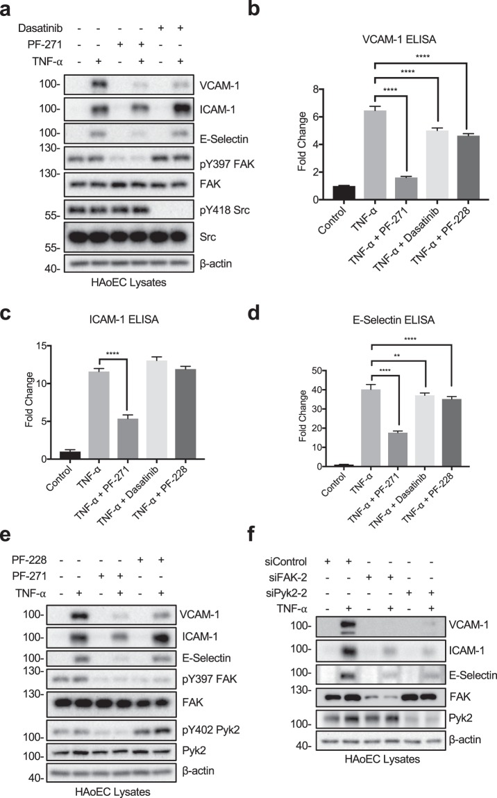
FAK/Pyk2 inhibition blocks TNF-α-induced pro-inflammatory adhesion molecule expression in HAoECs. (a) HAoECs were treated with DMSO, a dual FAK/Pyk2 inhibitor (PF-271, 2.5 μM) or a Src inhibitor (Dasatinib, 1 μM) for 1 h prior to TNF-α (10 ng/ml, 6 h) stimulation. Cropped images of immunoblotting for VCAM-1, ICAM-1, E-selectin, active FAK (pY397 FAK), FAK, active Src (pY418 Src), Src, and β-actin as loading control are shown. Full length blots shown in Supplemental Fig. 7. (b–d) HAoECs were treated with DMSO, a dual FAK/Pyk2 inhibitor (PF-271, 2.5 μM), a Src inhibitor (Dasatinib, 1 μM), or a FAK inhibitor (PF-228, 10 μM) for 1 h prior to TNF-α (10 ng/ml, 6 h) stimulation. Expression levels of (b) VCAM-1, (c) ICAM-1, and (d) E-selectin were determined using ELISA (n = 3, ±SEM). (e) HAoECs were treated with DMSO, a dual FAK/Pyk2 inhibitor (PF-271, 2.5 μM) or a FAK inhibitor (PF-228, 10 μM) for 1 h prior to TNF-α (10 ng/ml, 6 h) stimulation. Cropped images of immunoblotting for VCAM-1, ICAM-1, E-selectin, pY397 FAK, FAK, pY402 Pyk2, Pyk2, and β-actin as loading control are shown. Full length blots shown in Supplemental Fig. 8. (f) HAoECs were transfected with either control, FAK siRNA (400 pmol, 36 h), or Pyk2 siRNA (400 pmol, 36 h) prior to TNF-α (10 ng/ml, 4 h) stimulation. Cropped images of immunoblotting for VCAM-1, ICAM-1, E-selectin, FAK, Pyk2, and β-actin as loading control are shown. Full length blots shown in Supplemental Fig. 9. **p < 0.01, ****p < 0.0001.

