Figure 3.
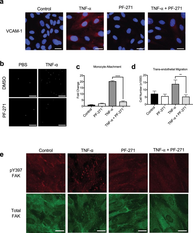
FAK/Pyk2 inhibition reduces TNF-α-induced in vitro inflammation. (a) HAoECs were treated with PF-271 (2.5 μM) for 1 h prior to TNF-α (10 ng/ml, 6 h) stimulation. Staining for VCAM-1 (red) and DAPI (blue) are shown. Scale bar (20 μm). (b-c) Monocyte adhesion assay. Primary mouse monocytes from bone marrow were labeled using Cell Tracker Green. HAoECs were treated with DMSO or PF-271 (2.5 μM) for 1 h prior to TNF-α (10 ng/ml, 6 h) stimulation. Monocytes were allowed to attach for 1 h prior to fixation. (b) Images of attached monocytes are shown. Scale bar (200 μm). (c) Attached monocytes were enumerated (n = 3, ±SEM). (d) HAoECs onto collagen I (10 μg/ml) coated Boyden chamber were treated with DMSO or PF-271 (2.5 μM) for 1 h prior to TNF-α (10 ng/ml, 6 h) stimulation. THP-1 cells were allowed to transmigrate the endothelial layer for 16 h. Transmigrated THP-1 cells were quantified (n = 3, ±SEM). (e) HAoECs were treated with PF-271 (2.5 μM) for 1 h prior to TNF-α (10 ng/ml, 6 h) stimulation. Staining of active FAK (pY397 FAK, red) or total FAK (green) are shown. Scale bar (20 μm). **p < 0.01, ****p < 0.0001.

