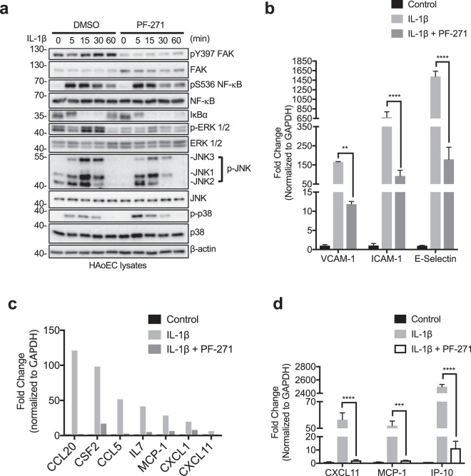
Figure 5.
FAK/Pyk2 activity promotes IL-1β-mediated pro-inflammatory molecule expression via transcriptional control. (a) HAoECs were treated with DMSO or PF-271 (2.5 μM) for 1 h prior to IL-1β (20 ng/ml) stimulation for 0 to 60 min. Cropped images of immunoblotting for pY397 FAK, FAK, active NF-κB (pS536 NF-κB), NF-κB, IκBα, active ERK (p-ERK), ERK, active JNK (p-JNK1, 2, and 3), JNK, active p38 (p-p38), p38, and β-actin as loading control are shown. Full length blots shown in Supplemental Fig. 13. (b) HAoECs were treated with DMSO or PF-271 (2.5 μM) for 1 h prior to IL-1β (20 ng/ml, 6 h) stimulation. RNA was collected, and RT-qPCR for cell adhesion molecules was performed (n = 3, ±SEM). (c) HAoECs were treated with DMSO or PF-271 (2.5 μM) for 1 h prior to IL-1β (20 ng/ml, 24 h) stimulation. RNA was collected, and RT-qPCR using an array for inflammatory cytokines, chemokines and their receptors. Shown are a selection of genes that are important in vascular inflammation and were reduced by PF-271 treatment upon IL-1β stimulation. (d) CXCL11, MCP-1, and IP-10 mRNA levels were verified by RT-qPCR (n = 3, ±SEM). **p < 0.01, ***p < 0.001, ****p < 0.0001.

