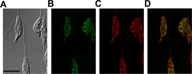
Figure 3.
Endoplasmic Reticulum (ER) localization of CPCT in L. major. Log phase promastigotes of cpct−/+GFP-CPCT were labeled with rabbit anti-T. brucei BiP antiserum followed by a goat anti-rabbit IgG-Texas Red antibody and subjected to confocal immunofluorescence microscopy. (A) Phase contrast; (B) GFP fluorescence; (C) Anti-BiP staining; (D) Merge of B and C. Scale bar: 5 µm. The overlap between BiP and GFP-CPCT was determined by the JaCOP Image J analysis of 30 cells (Table S1).

