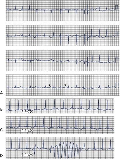
Figure 2: Twelve-lead ECG from a Patient with Hypokalaemia and Hypomagnesaemia.

A: Marked QTU prolongation and QTU alternans (arrowheads). B–D: Representative rhythm strips from the same patient showing tachycardia-dependent QTU alternans and torsade de pointes. Source: El-Sherif N, et al. 2011.4 Reproduced with permission from © Via Medica.
