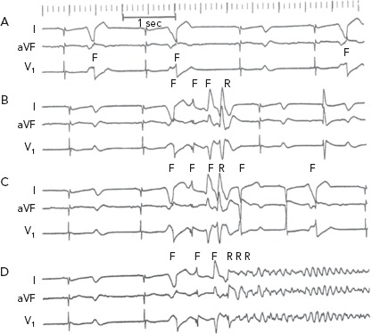
Figure 6: ECG Recordings from an In Vivo Canine Anthopleurin-A Surrogate Model of Long QT Syndrome Type 3.

The recordings are arranged chronologically, a few minutes apart. A: Stable bigeminal and trigeminal rhythm due to subendocardial discharge attributed to EAD-triggered activity from the same focus. This was followed several minutes later by runs of four- or five-beat polymorphic VT with remarkable repetition of the same QRS morphology (panels B and C). The first beat of each run arose from the same site of the bigeminal/ trigeminal beats in panel A. The second and third beats of each run arose from two different subendocardial focal sites; the fourth beat was reentrant in origin. The fifth beat in a five-beat run again was focal in origin and arose well after the end of the reentrant excitation and could be attributed to DAD-triggered activity. After approximately 10 minutes of repetitive non-sustained VT, the same three initial focal beats were followed by reentrant excitation that degenerated into ventricular fibrillation (VF) (panel D). F = focal discharge; R = reentrant excitation. Source: El-Sherif, 2001.84 Reproduced with permission from © Futura Publishing Company, Inc. 2001.
