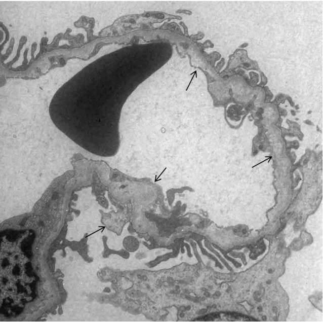
Fig. 1.

Electron microscopic examination of renal biopsy tissues in case 1. The ultrastructure of the glomerular basement membrane shows focal marked abnormalities with thickening, lysis, reticulation, and subepithelial protrusion of the lamina densa (arrow) (×8,000).
