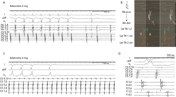
Figure 1: Responses of Atrial Tachycardias to Adenosine.

A: Focal atrial tachycardia from the lateral tricuspid annulus terminates with adenosine. B: In the same case as A, the electrogram at the successful ablation site shows a QS unipolar configuration with a sharp downstroke (on Lat TA 1 uni), which precedes other right atrial (RA) electrograms. RA electrograms are shown from a multipolar catheter looped around the lateral RA. C: Microreentrant tachycardia in a patient with prior ablation of AF. The tachycardia was localised to the anterior ridge between the left superior pulmonary vein and left atrial appendage. It was insensitive to adenosine. D: In the same case as C, multipolar Pentaray© catheter shows fragmented long-duration electrograms on two splines that encompass most of the tachycardia cycle length. CS = coronary sinus; dist = distal; lat = lateral; prox = proximal; TA = tricuspid annulus; uni = unipolar.
