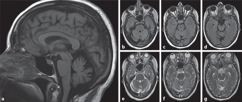
Fig. 4.
a–g MRI scans of a female heterozygous for NHE6 mutation at approximately age 51 show cerebellar atrophy; the individual was diagnosed with atypical parkinsonism. a The sagittal T1-weighted image demonstrates cerebellar atrophy. Axial FLAIR (b–d) and T2-weighted (e–g) images through the cerebellar hemispheres further highlight diffuse cerebellar volume atrophy.

