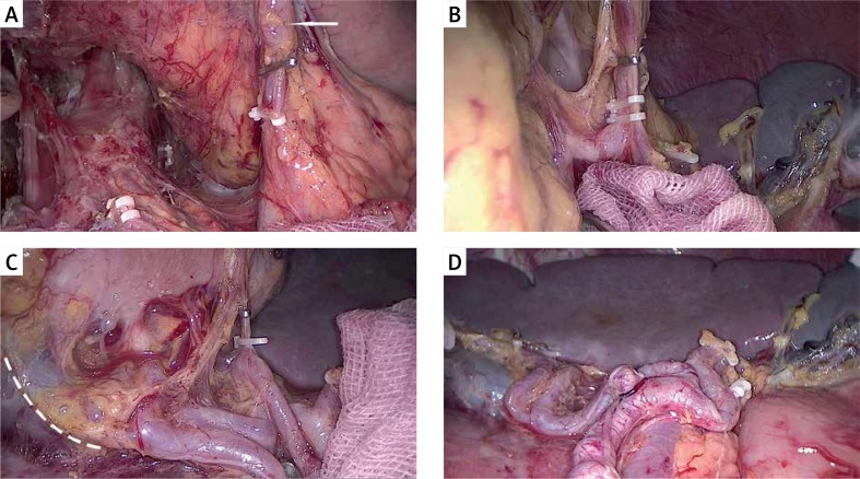
Photo 2.
An intraoperative image showing the procedures used to dissect LNs no. 10 and no. 11 along the splenic vessels. A – Exposing and clipping the posterior gastric artery (PGA). The arrow indicates the dissected no. 11 LN. B – Exposing and clipping the left gastroepiploic vessels (LGEVs). C – Exposing and clipping the short gastric arteries (SGA). The dashed line shows the partial left border of the “enjoyable space”. D – An overall view of the skeletonized splenic vessel trunk and branches

