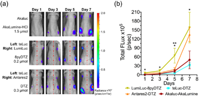
Figure 4. Tracking of tumor growth in a xenograft mouse model with various luciferase-luciferin pairs.

(a) BLI (n = 4) on day 1, 3, 5, and day 7. 104 luciferase-expressing HeLa cells were injected to the left and right dorsolateral trapezius regions and 105 cells were injected to the left and right dorsolateral thoracolumbar regions of NU/J mice. For i.v. administration of substrates, AkaLumine-HCl (1.5 μmol/mouse) and 8pyDTZ (0.2 μmol/mouse) were dissolved in normal saline, and DTZ (0.3 μmol/mouse) was formulated with a mixture of organic cosolvents. (b) Comparison of luciferase-luciferin pairs at tumor sites inoculated with 104 cells. (*p < 0.05 for LumiLuc-8pyDTZ and teLuc-DTZ, and for LumiLuc-8pyDTZ and Akaluc-AkaLumine; **p < 0.05 for LumiLuc-8pyDTZ and Antares2-DTZ).
