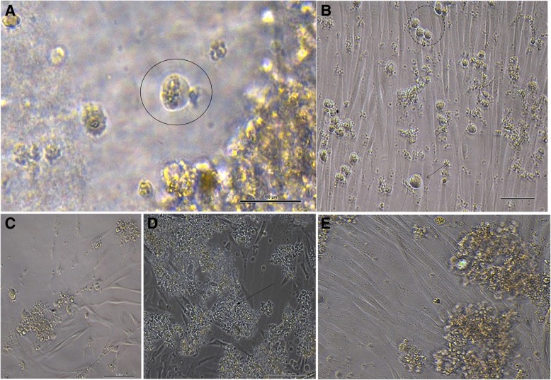
Fig. 1.
a Typical morphology of spermatogonia: rounded or oval cells and spiral nucleus related to the cytoplasm (circle). b, c Microscopic appearance of the formation of germ cell clumps (arrow) in small groups of cells (dotted circle) and continuous proliferation of clump-forming cells (asterisk) (2 days and 15 days after in vitro culture of cSSCs). d, e Formation of clumps by cells in suspension (arrow) among Sertoli cells (asterisk). (Scale bar = 100 μm)

